▶ 细胞重量变化研究
QPI技术能够对细胞微小的质量变化进行监控,具备的灵敏度。并且能够同时分析细胞的各种形态变化,诸如质量变化、面积、方向性等。这种对于大批量细胞的精确分析能力能够为肿瘤的起源和肿瘤耐药性的研究提供诸多帮助。
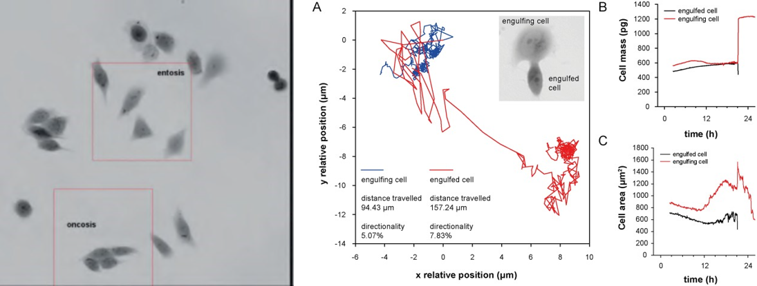
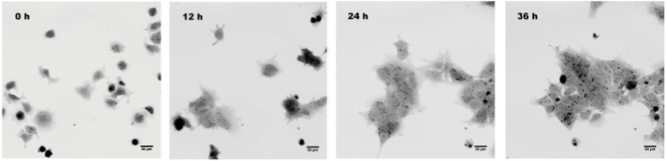
Role of entosis in oxidative stress resistance of PC-3 prostate cancer cells.
参考文献:Balvan J, Gumulec J, Raudenska M, Krizova A, Stepka P, Babula P, et al. (2015) Oxidative Stress Resistance in Metastatic Prostate Cancer: Renewal by Self-Eating. PLoS ONE 10(12): e0145016. https://doi.org/10.1371/journal.pone.0145016
▶ 干细胞长时间无标记成像及细胞周期研究
干细胞分化对于组织再生修复具有重要意义。为医学、干细胞治疗和发育生物学提供了许多新的研究方向。然而,传统的标记方案对于干细胞研究难免会对珍贵的干细胞造成不同程度的损伤。Q-Phase研究细胞时采用非入侵无标记的方式进行了采集,能够提供高速,高通量的细胞表征和分析。
Time-lapse differentiation of human embryonic stem cells. Samples provided by Dr. Jaroš, Faculty of Medicine, Masaryk University, Brno
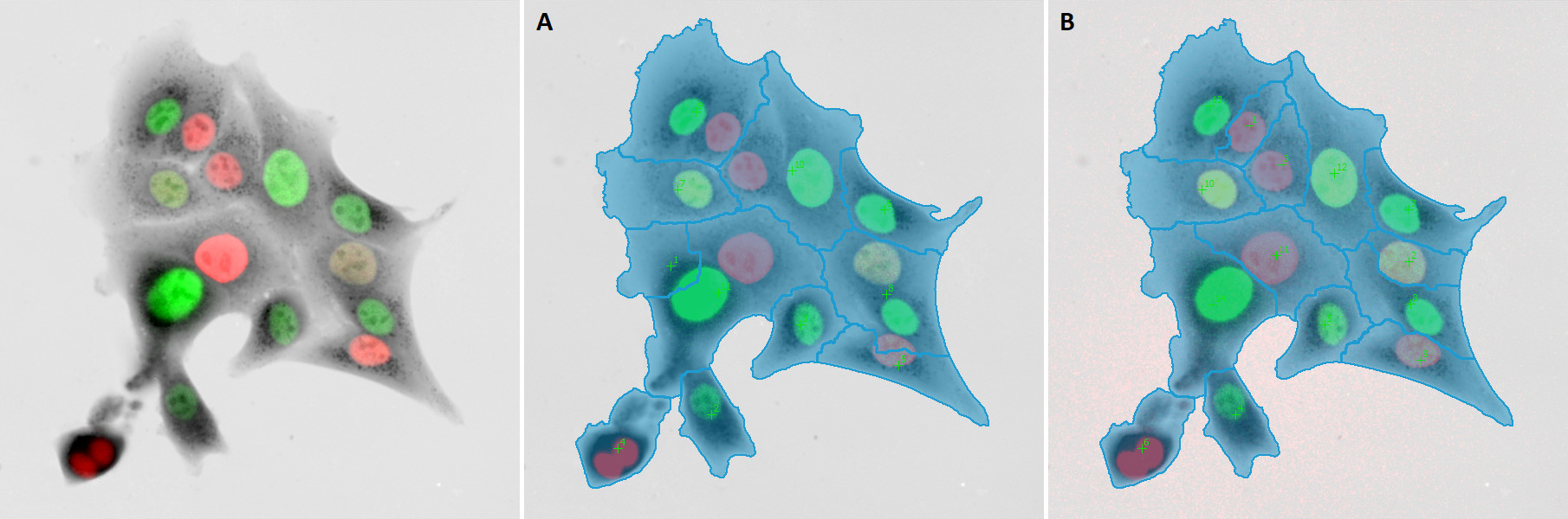
细胞周期的变化是细胞的基本特征。细胞周期的研究在传统上依靠对特定的标记或使用转基因系统,使得很难在不干扰细胞的情况下确定细胞周期阶段。Q-Phase独有的QPI模式能够在无标记的情况下监控细胞生长以及形态学和单细胞水平的表型变化。

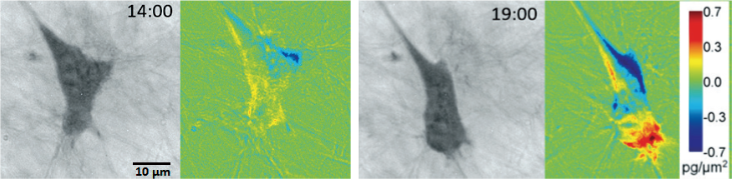
QPI images illustrating cell morphology at marked out points in the life cycle of LW13K2 cell
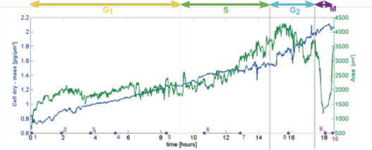
Changes in cellular mass and area during the cell cycle of LW13K2 cell. The value of mass has deen doubled between two mitosis.
▶ 精子的运动分析研究
精子计数测试能够分析人类精子的健康和活力。精子分析方法需要测量影响精子健康的三大因素:精子数量,精子的形状和运动。然而,精子细胞通常很获得标准显微图像。Q-Phase提供了一个快速可靠的精子细胞识别方法,从而便于快速评估精液中精子的数量和质量。

Semen analysis by Q-Phase system
▶ 在三维基质和不透明环境中成像:胶原基质中的细胞成像研究
三维环境中肿瘤细胞行为的观察与分析对于充分理解肿瘤侵袭性和转移形成具有十分重要的意义。然而,这样的实验在不使用特殊标记的情况下是很难的检测到的。通过Q-Phase所独有的QPI技术就能够使这一观察成为可能。癌细胞即使在分散的环境中,如三维胶原蛋白矩阵中也能够被清晰观测。
Migration of mesenchymal HT1080 cell within collagen matrix. Changes of mass distribution in migrating cell were analyzed by calculating the dynamic phase differences between consequent images.